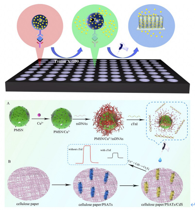
FIGURE 3.

(A) Scheme of the proposed liposomal PEC Immunoassay. Ab1 were first loaded into 96-well plates, and then samples were incubated with Ab1 for 1 h. The QDLL-Ab2 was introduced after the immunorecognition of cTnI. Triton X-100 (10%) was introduced to release the QDs that were later captured by TiO2-NTs electrode. The photocurrents were generated due to the sensitization effect (Xue et al., 2019). Copyright 2020 American Chemical Society. (B) Schematic principle of cTnI detection using PMSN/Cu2+/ssDNAs. Positive-charged PMSNs were added into Cu2+ solutions. Cu2+ entered the pores of PMSNs via diffusion. Later, ssDNAs bound cTnI specifically was added and attached on the surface of PMSNs. Once the cTnI was recognized by ssDNAs, the complexes reacted with CdS that was functionalized onto the PSATs, to form CuxS and diseased the photocurrent signal. The changes of photocurrent were correlated to the concentration of cTnI (Gao et al., 2019). Copyright 2020 from Elsevier B.V.
