
Fig. 2.
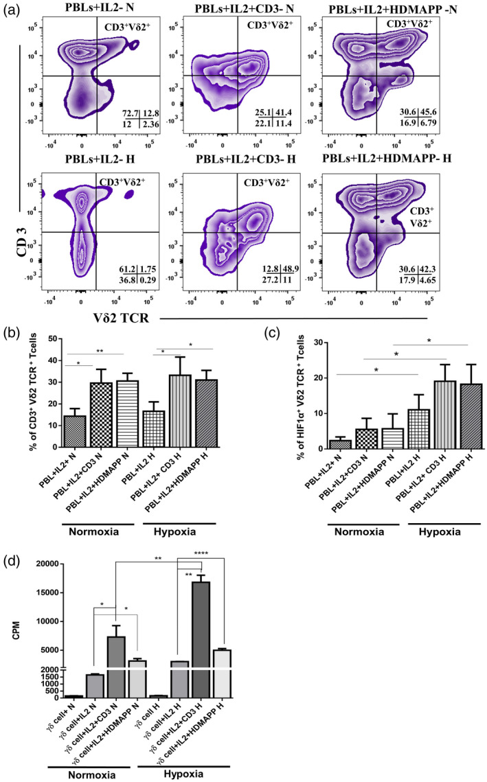
Expansion of gamma delta T (γδT) cells from peripheral blood lymphocytes (PBLs) upon T cell receptor (TCR) stimulation is unaltered under hypoxia. Healthy individuals’ PBLs (HI‐PBL) (n = 5) were expanded with recombinant human interleukin (rhIL‐2) alone, rhIL‐2 + αCD3 or rhIL‐2 + 1‐hydroxy‐2‐methyl‐2‐buten‐4‐yl 4‐diphosphate (HDMAPP) for 12 days under hypoxia (H) or normoxia (N), and assessed for expression of CD3+ Vδ2 TCR+ phenotype (a,b) and hypoxia‐inducible factor 1α (HIF1α) (c). (a) The representative figure of CD3+ Vδ2 TCR+ cells in stimulated HI‐PBLs gating was performed on lymphocytes. The values shown in the figure indicate the percentage‐positive population. (b) Mean ± standard error of the mean (s.e.m.) of the percentage of percentage‐positive CD3+ Vδ2 TCR+cells in HI‐PBLs. (c) Mean ± s.e.m. of the percentage of positive HIF1α+ Vδ2 TCR+cells in HI‐PBLs (gated on CD3+ Vδ2 TCR+ cells; *P < 0·05; **P < 0·01). (d) γδT cells isolated from HI‐PBLs by magnetic‐activated cell sorting (MACS) (n = 3) were stimulated with rhIL‐2, rhIL‐2 + αCD3 or rhIL‐2 + HDMAPP or kept unstimulated and cultured for 72 h in H or N. The proliferation of γδT cells was assessed by [3H]‐thymidine incorporation assay. Results indicated are mean ± s.e.m. of three independent experiments (*P < 0·05; **P < 0·01; ****P < 0·005).