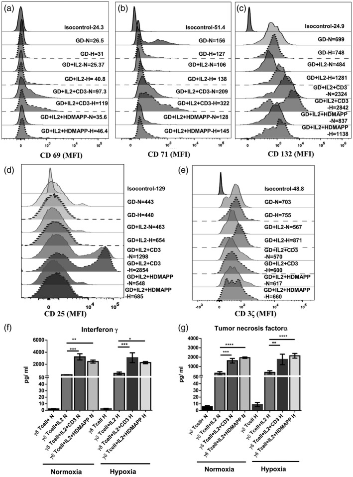
Fig. 3.

Hypoxia does not alter the activation status and the cytokine secretion of gamma delta T (γδT) cells. γδT cells isolated from healthy individuals’ peripheral blood lymphocytes (HI‐PBL) (n = 3) were stimulated with recombinant human interleukin (rhIL‐2), rhIL‐2+αCD3 or rhIL‐2+1‐hydroxy‐2‐methyl‐2‐buten‐4‐yl 4‐diphosphate (HDMAPP) or kept unstimulated and cultured for 72 h in hypoxia (H) or normoxia (N). The mean fluorescence intensity (MFI) of activation markers (a) CD69, (b) CD71, (c) CD132, (d) CD25 and (e) CD3ζ was assessed using multi‐color flow cytometry. Gating was performed on CD3+ Vδ2 TCR+ T cells. (f,g) γδT cells isolated from HI‐PBLs (n = 3) were stimulated with rhIL‐2, rhIL‐2 + αCD3 or rhIL‐2 + HDMAPP and cultured for 24 h in H or N. The cell‐free supernatants were collected and assessed for secreted cytokines (f) interferon (IFN)‐γ and (g) tumor necrosis factor (TNF)‐α using a cytometric bead array (CBA) kit. All the results indicated are mean ± standard error of the mean (s.e.m.) of three independent experiments (*P < 0·05; **P < 0·01; ***P < 0·005; ****P < 0·001).
