
Fig. 6.
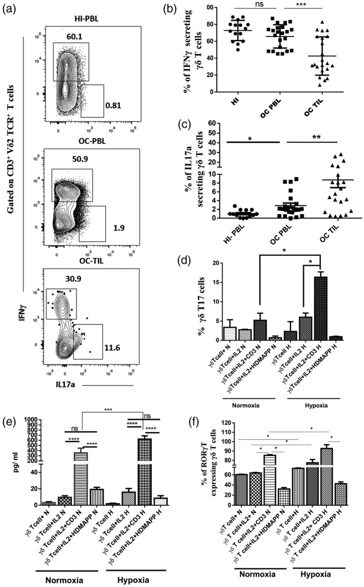
Gamma delta T17+ [γδT cells that produce interleukin (IL)‐17] cells are enhanced in the presence of hypoxia. (a) Representative figure showing the expression of interferon (IFN)‐γ and IL‐17a gated on CD3+ Vδ2 T cell receptor (TCR)+ γδT cells in healthy individuals’ peripheral blood lymphocytes (HI‐PBLs) and PBLs and tumor‐infiltrating lymphocytes (TILs) (paired samples) of the oral cancer (OC) patient. The values shown in the figure indicate the percentage‐positive population. (b,c) Intracellular expression of (b) IFN‐γ and (c) IL‐17a was assessed on a CD3+ Vδ2 TCR+ subset of γδT cells in HI‐PBLs (n = 15) and OC patient paired samples (n = 22) PBLs and TILs. (d,e) γδT cells isolated from HI‐PBLs were stimulated with recombinant human interleukin (rhIL‐2), rhIL‐2 + αCD3 or rhIL‐2 + 1‐hydroxy‐2‐methyl‐2‐buten‐4‐yl 4‐diphosphate (HDMAPP) or kept unstimulated and cultured for 72 h in hypoxia (H) and normoxia (N). γδT cells were assessed for intracellular expression of IL‐17a using multi‐color flow cytometry. (d) Mean ± standard error of the mean (s.e.m.) of the percentage of positive γδT17+ cells and (e) γδT cells were stimulated with rhIL‐2, rhIL‐2 + αCD3 or rhIL‐2 + HDMAPP and cultured for 24 h in the presence or absence of H. The cell‐free supernatants were collected and assessed for the secretion of IL‐17a (pg/ml) using a cytometric bead array (CBA) kit. (f) γδT cells were assessed for intracellular expression of RAR‐related orphan receptor gamma (RORγt) using multi‐color flow cytometry, mean ± s.e.m. of the percentage of positive RORγt+ γδT cells using multi‐color flow cytometry. All the results indicated are mean ± s.e.m. of three independent experiments (*P < 0·05; **P < 0·01; ***P < 0·005; ****P < 0·001).