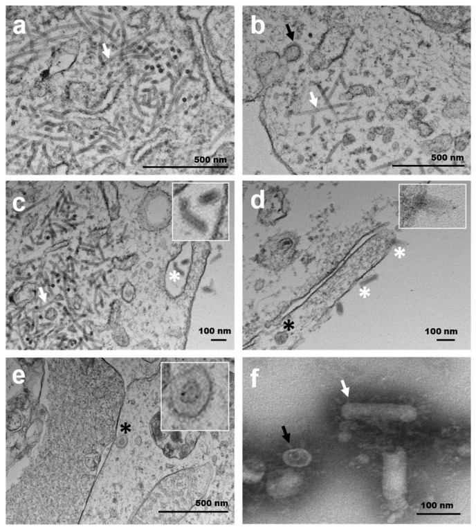
Figure 5.
Transmission electron micrographs of FHM (a,b) and E11 cells (c–f) cells inoculated with chinook salmon bafinivirus Cefas-W054 isolate. (a–c) Spherical (black arrows) and bacillary (white arrows) viral nucleocapsids were observed within the cytoplasm of these cells. (c–e) Mature enveloped spherical (black asterisks) and bacillary virions (white asterisks). Top inserts show the detail of mature virions with spikes on the surface. (f) Transmission electron (negative staining) of purified virions from the E11 cell supernatant. The external surface of spikes can be seen surrounding spherical (black arrows) and rod-shaped (white arrows) virions.

