Figure 6.
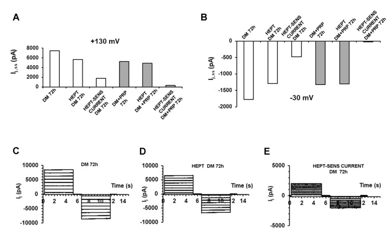
Evaluation of the current flowing through the GJs by acute addition of heptanol under different experimental conditions. (A,B) Evaluation of Ij,ss (pA) related to a typical cell pair cultured for 72 h in differentiation medium (DM, white bars on the left side of each graph) or to a cell pair cultured for 72 h in DM + PRP (grey bars on the right). For clarity, only the current amplitude values obtained in response to a representative voltage pulse to +130 mV is reported in A, and to −30 mV in B. The current value recorded from the myofibroblast pair (DM 72 h) resulted clearly reduced after the acute addition of heptanol (HEPT, 1 mM) to the bath solution (HEPT DM 72 h). The current flowing through the GJs at the steady-state (HEPT-SENS CURRENT DM 72 h) is obtained by subtracting the current values recorded in the presence of heptanol from those recorded in DM alone. In contrast, the current amplitude recorded from the cell pair cultured in DM + PRP after acute addition of heptanol (HEPT DM + PRP 72 h) showed a slight reduction compared to DM + PRP 72h, having the heptanol-sensitive current a very small value (HEPT-SENS CURRENT DM + PRP 72h). Similar results were systematically observed for any voltage step applied in the bulk of the cell pairs investigated, suggesting a very small amount of functional GJs expressed under PRP treatment. (C) Characteristic time course of Ij (pA) recorded from a cell pair cultured in DM (DM 72 h) that exceptionally showed voltage independent features. (D) The same current traces recorded after acute heptanol addition (HEPT DM 72 h). (E) Resulting current traces (HEPT-SENS CURRENT DM 72 h) obtained by subtracting currents in D from currents in C, and representing the small heptanol-sensitive flux through the GJs. Note the different ordinate scale in E.

