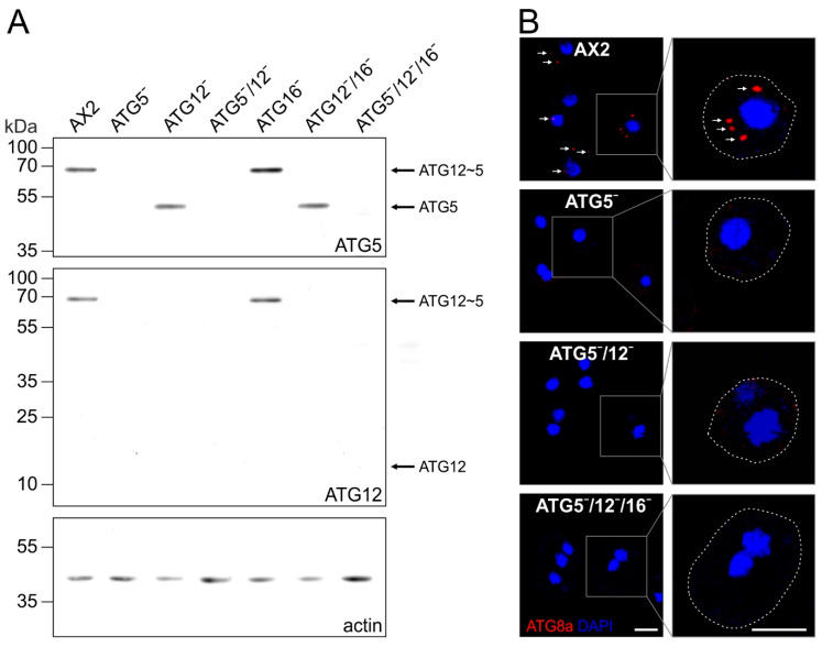
Figure 3.
Verification of the different mutant strains by immunoblotting and immunofluorescence analysis of ATG8a-positive autophagosomes. (A) Immunoblotting of total cell lysates of wild-type AX2, ATG5¯, ATG12¯, ATG5¯/12¯, ATG16¯, ATG12¯/16¯, and ATG5¯/12¯/16¯ cells. The ATG12~5 conjugate was detected at about 68 kDa in AX2 and ATG16¯ cell lysates but not in atg5 and atg12 knock-out strains. Unconjugated ATG5 of about 46 kDa was detected in ATG12¯ and ATG12¯/16¯ cells. No unconjugated ATG12 of about 14 kDa was detectable. Actin was used as a loading control. ATG5, ATG12 and actin were visualised on the same membrane. Top row, ATG5 pAb; middle row, ATG12 mAb; bottom row, actin mAb. (B) Immunofluorescence microscopy of AX2, ATG5¯, ATG5¯/12¯ and ATG5¯/12¯/16¯ cells. Cells were fixed with cold methanol and stained with the ATG8a pAb. Puncta representing ATG8a-positive autophagosomes (arrows) were only detected in AX2 wild-type cells. Cell boundaries in the enlarged insets are indicated by dotted lines. Nuclei were visualised by DAPI staining. Scale bar, 5 µm.

