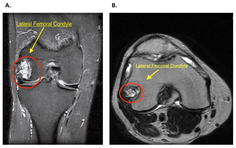
Figure 1.
(A) Right knee magnetic resonance image, fat-saturated FSE-IR (fast spin-echo inversion-recovery). Coronal plane shows a multiloculated cystic area (red circle) in the lateral femoral condyle with proximal bone marrow edema. (B) Right knee magnetic resonance image, non-fat-saturated T2 weighted. Axial plane image shows the cystic area (red circle) used for localization and planning of procedure.

