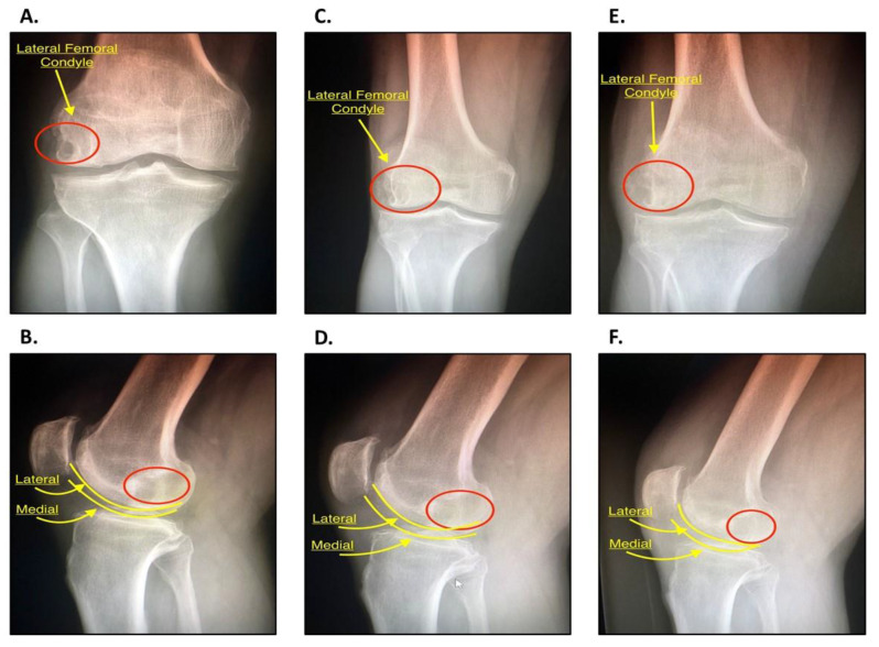
Figure 6.
(A) Anteroposterior (AP) radiograph of the right knee, pre-operative. The lateral femoral condyle can be visualized with the area of decreased opacity representing the SC (red circle). The joint space is well preserved with mild patellofemoral OA with osteophytes. (B) Lateral radiograph of the right knee, pre-operative. The red circle depicts the same SC. The lateral and medial femoral condyles are outlined in yellow. Both (A) and (B) will be used as a baseline for comparison. (C) AP radiograph of the right knee 3 months following IOBP. The lateral femoral condyle can be visualized with signs that the area of the previous SC is filling (red circle). (D) Lateral radiograph of right knee 3 months following operation. The red circle depicts the area of the SC. (E) AP radiograph of the right knee 6 months following IOBP. The area of the previous SC (red circle) has increased in opacity in comparison to the previous image at 3 months. (F) Lateral radiograph of the right knee six months following IOBP. The area of the previous SC (red circle) is more opaque, an indication that there is progressing filling of the previous lesion. This suggest that the IOBP has been successful.

