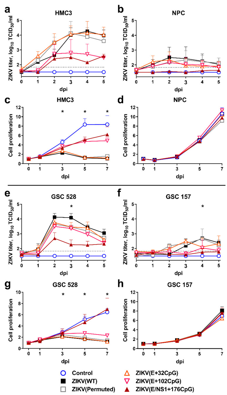
Figure 2.
Infection kinetics in nonmalignant human brain cells (HMC3 (a) and NPC (b)) and tumor glioblastoma stem cells (GSC 528 (e) and GSC 157 (f)) after inoculation at multiplicity of infection (MOI) of 0.01. Cell culture supernatants in 96-well plates were collected and viral titers were measured using the endpoint dilution assay. The dotted line represents the limit of detection. Cell proliferation assay after inoculation of cells (HMC3 (c) and NPC (d), GSC 528 (g), and GSC 157 (h)) with MOI of 1. Whiskers represent the standard error of the mean (SE) from three biologically independent replicates with three technical replicates. “dpi”—days post-inoculation. The asterisk (*) indicates p < 0.05 vs. WT (a,b,e,f) and control (c,d,g,h): (c) WT and E+32CpG at 3–7 dpi, permuted control at 5–7 dpi; (e) E/NS1+176CpG at 3 dpi; (f) E+32CpG and E/NS1+176CpG at 4 dpi; (g) WT, permuted control, E+102CpG at 3–7 dpi.

