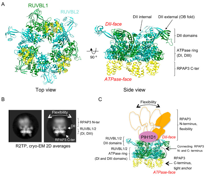
Figure 2.
Structure of RUVBL1-RUVBL2 AAA-ATPases and the R2TP complex. (A) Top and side view of the atomic structure of the core of the human R2TP complex comprising one RUVBL1-RUVBL2 hexameric ring bound to three copies of the RPAP3 C-terminal domain [37]. Subunits, domains, and regions are indicated. (B) Two representative 2-D averages of cryo-EM images obtained for the R2TP complex [37]. Domains of RUVBL1-RUVBL2 and RPAP3 are indicated. (C) Cartoon representing the location of PIH1D1 and RPAP3 N-terminal end at the DII-face of the RUVBL1-RUVBL2 ring, and the flexibility of RPAP3 is indicated.

