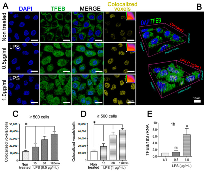
Figure 4.
Purified LPS from A. actinomycetemcomitans induces TFEB activation. OKF6/TERT-2 cells were incubated with 0.5 or 1 µg/mL of purified LPS from A.a (serotype b) and analyzed by (A) immunofluorescence visualized by confocal microscopy. Blue: DNA rich structures strained with DAPI; green: transcription factor EB labeled with anti-TFEB antibody; merge: digital overlap of blue and green channels; yellow: colocalized voxels from merged file. (B) 3-D reconstruction of confocal image data showing evident TFEB-labeling in LPS-stimulated JEK nuclei, scale bar 10 µm. Quantification of colocalized voxels of LPS-stimulated cells at different time points. (C,D) Quantification of colocalized voxels from DAPI and TFEB channels in LPS-stimulated cells at different time points. Cells were stimulated with 0.5 (C) or 1 µg/mL (D) of LPS for the indicated period of time. (E) Quantitative real-time PCR of TFEB transcripts abundance normalized to 18S rRNA levels after 1 h of LPS stimulation. * p < 0.05.

