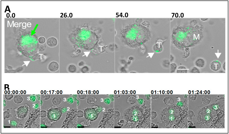
Figure 2.
HIV-1 transfer between macrophages and CD4+ T cells. (A) HIV-1 infected macrophages transfer GFP-HIV-1 to a CD4+ T cell that is scanning the macrophage surface via transient adhesive interactions (taken from [56]). The green arrow indicates the virus-containing compartment. The white arrows show viral particles engaged with the T cell surface. Numbers above images represent minutes:seconds after initiating coculture. Scale bar = 10 μm. (B) HIV-1-GFP infected CD4+ T cells being engulfed by a macrophage (taken from [65]). Numbers above images represent hours:minutes:seconds. Scale bar = 10 μm.

