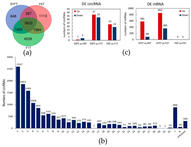
Figure 1.
The novel detected circRNAs and differentially expressed analysis in theca cells: (a) Venn diagram of novel detected circRNAs in three types of theca cells; (b) distribution of circRNAs on the chromosome; and (c) the number of differentially expressed circRNAs and mRNAs. SYF: small yellow follicle, F6: smallest hierarchical follicle, F1: largest hierarchical follicle. T: theca cells.

