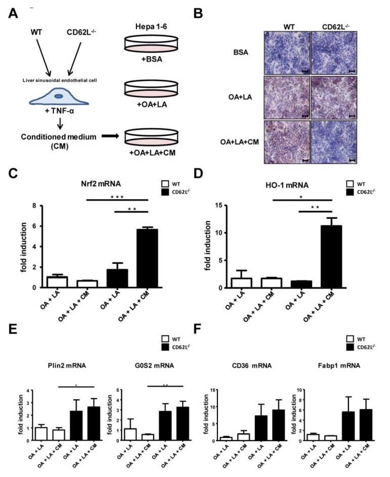
Figure 5.
Activation of the anti-oxidative stress response and increased fat turnover in Hepa1-6 cells after stimulation with medium of activated liver sinusoidal endothelial cells (LSEC) from CD62L−/− mice. (A) Isolated primary LSEC of WT and CD62L−/− mice were activated by stimulation with bovine serum albumin (BSA) (control) or TNF-α for 24 h. Hepa1-6 cells were stimulated with linoleic acid-oleic acid-albumin (OA + LA) and additionally with conditioned medium (CM) (OA + LA + CM) of TNF-α-stimulated primary LSEC of WT and CD62L−/− mice for 24 h. (B) Representative Oil Red O-stained tissue culture slides are depicted showing the progressive decrease of Oil Red O-staining in CD62L−/− mice after stimulation with OA + LA + CM (400×). (C) Nrf2, (D) HO-1, (E) Plin2, G0S2, (F) CD36 and Fabp1 mRNA expression was analysed in the stimulated Hepa1-6 cells via RT-qPCR. For quantification values are expressed as fold induction over the mean values obtained for OA + LA treated Hepa1-6 cells (* p < 0.05, ** p < 0.01, and *** p < 0.001).

