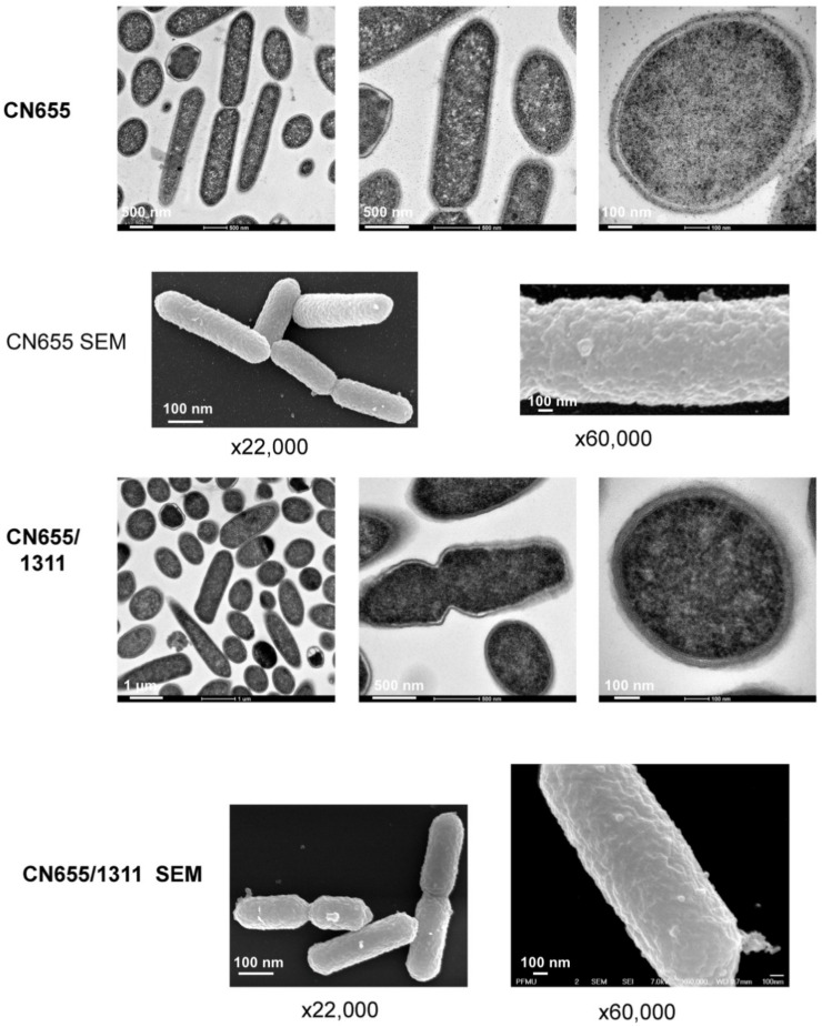
Figure 8.
Ultrastructural morphology of CN655 and CN655/p1311. Bacteria from 18 h TGY culture were processed for transmission electron microscopy and scanning electron microscopy (SEM). CN655 showed well-delineated bacterial wall layers, whereas the bacterial wall of CN655/p1311 was disorganized with diffuse and enlarged wall layers. CN655/p1311 showed more abundant blebbings on the bacterial surface. About 100 bacterial cells were observed for each preparation.

