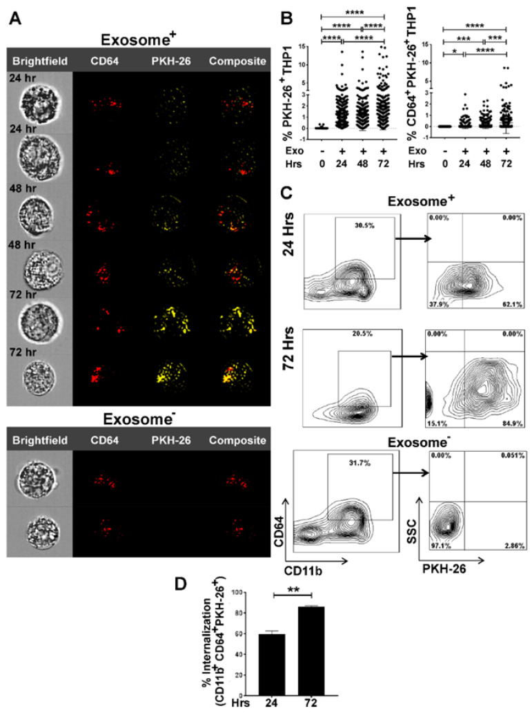
Figure 2.
ImageStream and flow cytometry analyses quantify time-dependent internalization of tumor cell-derived exosomes by THP-1 cells. THP-1 cells were seeded and differentiated into M0 macrophages upon overnight stimulation with PMA (20–100 ng/mL). M0 macrophages were then co-cultured with PKH-26 stained A549-derived exosomes in 10:1 ratio (10 exosomes/cell) for 24 to 72 h. Image Stream analysis showing Bright field, CD64+, PKH-26+, and composite images. (A) Time-dependent internalization of exosomes by CD64+ populations assessed by internalization of PKH-26 stained A549-derived exosomes. CD64+ population, without internalization indicates M0 phenotype. (B) MATLAB analysis of percentage of PKH-26+ and CD64+PKH-26+ signals to show time-dependent internalization of exosomes. Fluorescent signals were collected from 300 cells for each time point. (C) Representative flow cytometry of exosome internalization analysis at 24 h and 72 h. Group comparisons of 24 h exosome-, 24 h exosome+, and 72h exosome+ were made. After co-culture, cells were prepared for flow cytometry and stained with CD64, CD11b, and PKH-26. The experiment was repeated twice, with three replicates per sample in each experiment. Exosome- sample was used as control (D) Percentage of internalization by THP-1 macrophages as CD11b+CD64+PKH-26+ showing a significant increase of uptake in 72 h compared to 24 h * p < 0.05, ** p < 0.01, *** p < 0.001, **** p < 0.0001.

