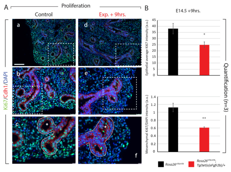
Figure 3.

Analysis of proliferation in E14.5 lungs after 9 h of Fgfr2b inhibition. (A) Control (a–c) and experimental (d–f) lungs were stained for Ki67 and Cdh1 to assess proliferation in the epithelial and mesenchymal compartments (separated by the white line in c and f). Note the multilayered and disorganized epithelium invading the lumen of experimental lung buds (compare asterisks in c and f), which is a hallmark of Fgfr2b inhibition at E12.5 as well [13]. Scale bar: (a, d) 100 µm, (b, e) 50 µm, (c, f) 20 µm. (B) Quantification of Ki67 expression showing a significant decrease in proliferation in both the epithelial and mesenchymal compartments. (a.u. = arbitrary units) (* p-value < 0.05, ** p-value < 0.01).
