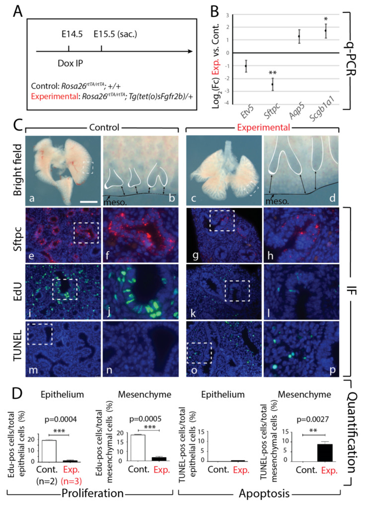
Figure 4.

Effects of transient Fgfr2b signaling inhibition for 24 h in E14.5 lungs. (A) Experimental design: pregnant females carrying experimental and littermate control embryos were injected with a single Dox-IP and sacrificed 24 h later. (B) qPCR analysis showing downregulated expression of AT2 markers Etv5 (not significant) and Sftpc (n = 3; ** p < 0.01), and upregulated expression of the AT1 marker Aqp5 (not significant), and the proximal epithelial marker Scgb1a1 (n = 3; * p < 0.05). (C) Brightfield (a–d) and immunofluorescent (e–p) images of control and experimental lungs. Note the elongated epithelial tubes, reduced distal tip branching, and increased distance between tips and mesothelium (black arrows) in experimental lungs compared to controls (d vs. b). Staining for Sftpc shows a decrease in the epithelium of experimental lungs (h vs. f), confirming the qPCR expression data. Staining for EdU and TUNEL to assess proliferation and apoptosis, respectively, showed decreased proliferation in both the epithelial and mesenchymal compartments of experimental lungs compared to controls (l vs. j), while apoptosis was increased only in the mesenchyme of experimental lungs (p vs. n). (meso. = mesothelium). Scale bar: (a, c) 750 µm, (b, d) 95 µm, (e, g, i, k, m, o) 75 µm, (f, h, j, l, n, p) 30 µm. (D) Quantification of EdU and TUNEL expressions in the epithelial and mesenchymal compartments of experimental vs. control lungs. (** p-value < 0.01, *** p-value < 0.0001).
