Figure 5.

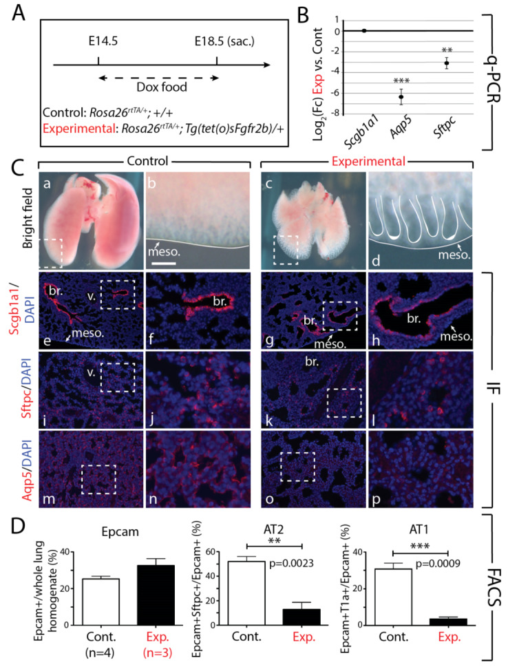
Continuous inhibition of Fgfr2b signaling from E14.5 to E18.5 leads to impaired AT1 and AT2 formation. (A) Experimental design: pregnant females carrying experimental and littermate control embryos were fed Dox food continuously for four days before being sacrificed at E18.5. (B) qPCR analysis showing significant downregulation of the AT1 marker Aqp5 and the AT2 marker Sftpc in experimental versus control lungs. Scgb1a1 shows no change in expression. (n = 3; ** p-value < 0.01, *** p-value < 0.001). (C) Brightfield (a–d) and immunofluorescent (e–p) images of control and experimental lungs. Note the smaller overall size and the dilated epithelial branches of experimental lungs compared to controls (c and d vs. a and b). Staining for Scgb1a1 shows a clear boundary between Scgb1a1+ proximal and Scgb1a1-negative distal epithelium in control lungs (e and f), whereas an ectopic distribution is seen in experimental lungs (g and h), with Scgb1a1+ cells being found in distal regions of the lung. Staining for Sftpc (i-l) and Aqp5 (m-p) reveal a downregulation of these proteins in experimental compared to control lungs, confirming the downregulation of these genes seen in the qPCR analysis (B). (meso. = mesothelium, br. = bronchus, v. = vessel). Scale bar: (a, c) 750 µm, (b, d) 95 µm, (e, g, i, k, m, o) 150 µm, (f, h, j, l, n, p) 30 µm. (D) FACS-based quantification shows no significant difference in the proportion of Epcam+ (epithelial) cells relative to the total (CD45-CD31-) lung cells in experimental versus control lungs, but a significant decrease in the proportion of AT2 (Epcam+ Sftpc+ from total Epcam+) (** p-value < 0.01) and AT1 (Epcam+T1alpha+ from total Epcam+) (*** p-value < 0.001) cells in experimental versus control lungs.
