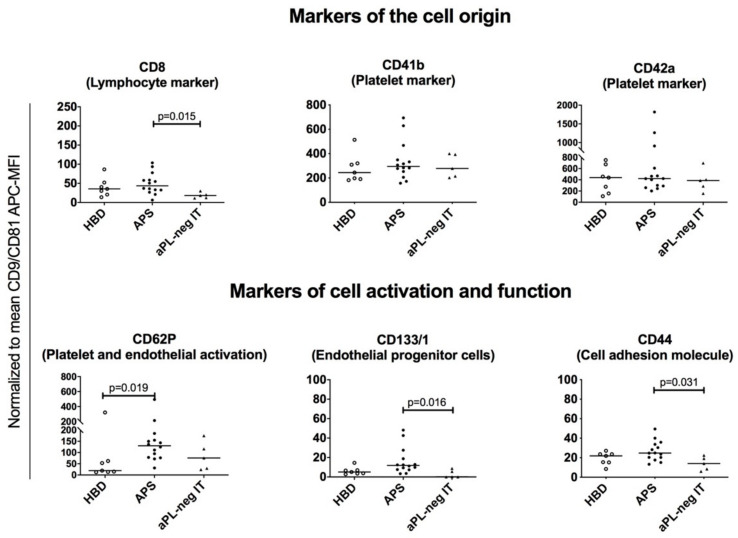
Figure 4.
Normalized median fluorescence intensities (MFI) of the surface protein profiles of the plasma-derived sEVs from healthy blood donors (HBD), patients with antiphospholipid syndrome (APS) and aPL-neg patients with idiopathic thrombosis (aPL-neg IT). The nonparametric Kruskal–Wallis test with Dunn’s multiple comparison adjustment was used. Grouped surface protein profiles are shown indicating the cell of origin (upper panel) and cell activation status/functional properties of the sEVs (lower panel).

