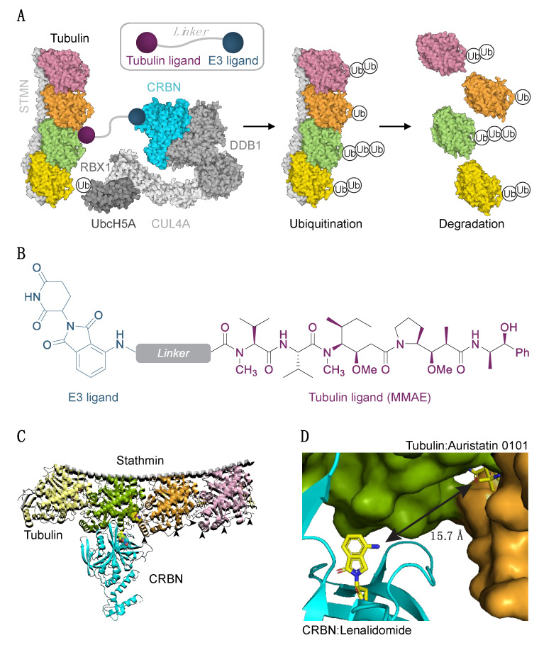
Figure 1.
Experimental strategy to degrade tubulin using the PROTAC technology. (A) Schematic diagram of protein degradation strategy. Recruitment of the E3 ligase to tubulin heterodimer via the tubulin/E3 ligand molecule would cause tubulin ubiquitination (Ub) and subsequent proteasome-mediated degradation. Structure of two tubulin heterodimers (pink, orange, green, yellow) bound to stathmin (STMN, PDB ID 4x1i) and the CRL4CRBN E3 ligase complex is shown (RBX1/CUL4/DDB1, PDB ID 4a0K; CRBN PDB ID 5fqd; UbcH5A, PDB ID 2c4p). (B) Structure of a chimeric compound. (C) Docking pose of CRBN and tubulin. Arrowheads indicate some of the exposed lysine residues in tubulins. (D) The shortest pairwise distance between lenalidomide and auristatin-0101 from pose in (C) is permissive of the degrader approach.

