Figure 3.
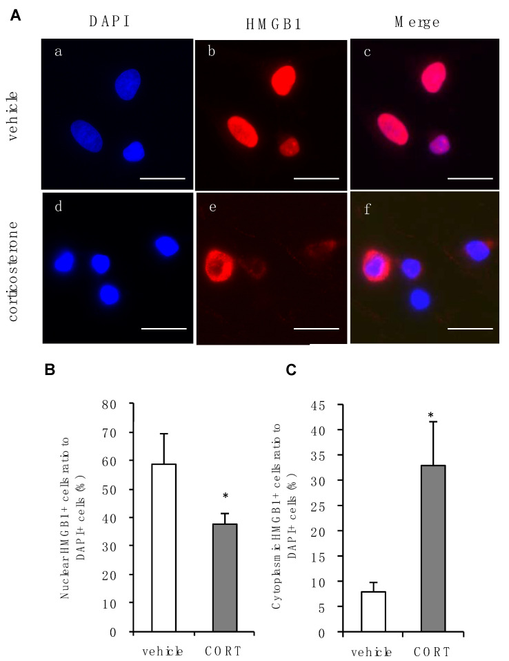
Effects of corticosterone on intracellular localization of HMGB1 in primary cultured cortical astrocytes. (A) Immunocytochemistry of HMGB1 in rat primary cultured cortical astrocytes. Corticosterone-treated cells (CORT; 1 μM, 24 h) exhibited subcellular HMGB1 expression compared with vehicle. Images a, b and c, or d, e and f, are from the same fields, respectively. Scale bar = 20 μm. (B) The percentage of nuclear staining of HMGB1 to total DAPI positive cells decreased after corticosterone treatment. The data are expressed as the mean ± SEM. * p < 0.05 vs. vehicle (t-test; n = 3–4). (C) The percentage of cytoplasmic staining of HMGB1 to total DAPI positive cells increased after corticosterone treatment. The data are expressed as the mean ± SEM. * p < 0.05 vs. vehicle (t-test; n = 3–4).

