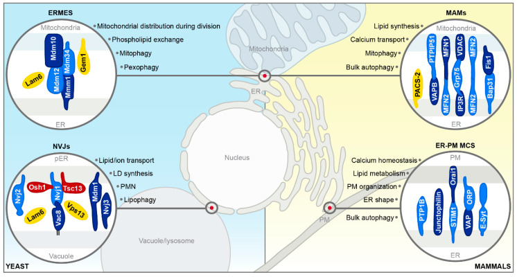
Figure 2.
Schematic overview of selected membrane contact sites. Tether proteins (blue), proteins with other functions (red) and regulatory proteins (yellow) are depicted. For a general description of membrane contact sites (MCS) in yeast (blue) and mammals (yellow), please see main text. ER = endoplasmic reticulum; ERMES = ER–mitochondria encounter structure; pER = perinuclear ER; NVJs = nucleus–vacuole junctions; LD = lipid droplet; PMN = piecemeal microautophagy of the nucleus; MAMs = mitochondria-associated membranes; PM = plasma membrane.

