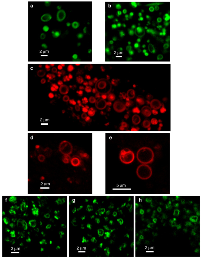
Figure 3.
Lysosomal membrane staining with naturally fluorescent lysosomotropic compounds. Vacuolin-1-treated U2OS cells were loaded for 45 min with LysoTracker Green DND-26 (LTG) (a,b), daunorubicin (DNR) (c–e) or nintedanib (NTD) (f–h) and imaged with a confocal Zeiss LSM 710 microscope (×63 magnification). All images are representative of data collected from at least three independent experiments. See also Figure S4.

