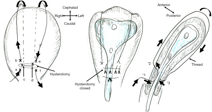
Figure 1.
Schematic presentation of B-Lynch uterine compression suture and intrauterine stent placement. A thread compresses the uterine lumen in a ventral-caudal (anterior-posterior) direction, thereby achieving hemostasis of postpartum hemorrhage. The marks (x: a-f) indicate the place where the needle penetrates the anterior (a, b, e, and f) and posterior (c and d) uterine wall. As * indicates, the needle penetrates the anterior (*1, *2: a, b, e, and f) and posterior (*3: c and d) uterine walls but does not penetrate the anterior-posterior uterine wall. A new intrauterine stent can be placed intrauterine, thereby preventing intrauterine adhesion, one of the most important adverse events of B-Lynch suture.

