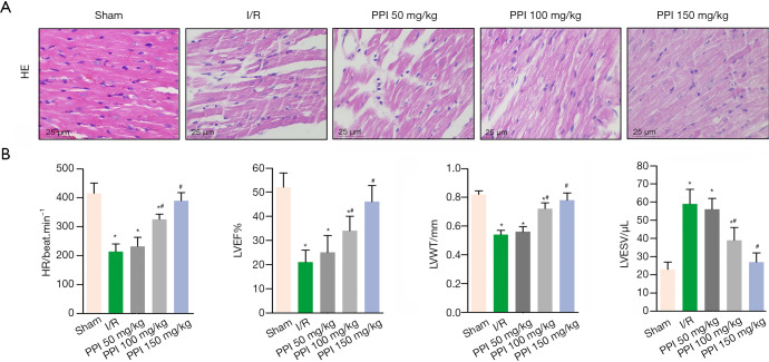
Figure 1.
PPI prevented cardiac dysfunction. (A) After 24 h of reperfusion, myocardial histology was detected by HE staining; representative images were taken under 200× magnification. (B) Left ventricular ejection fraction (LVEF), left ventricular wall thickness (LVWT) and left ventricular end-systolic volume (LVESV) were assessed by echocardiography. Data are presented as the mean ± SD (n=10). *, P<0.05 vs. control group, #, P<0.05 vs. I/R group. All independent experiments were repeated at least three times. PPI, polyphyllin I; I/R, ischemia/reperfusion.

