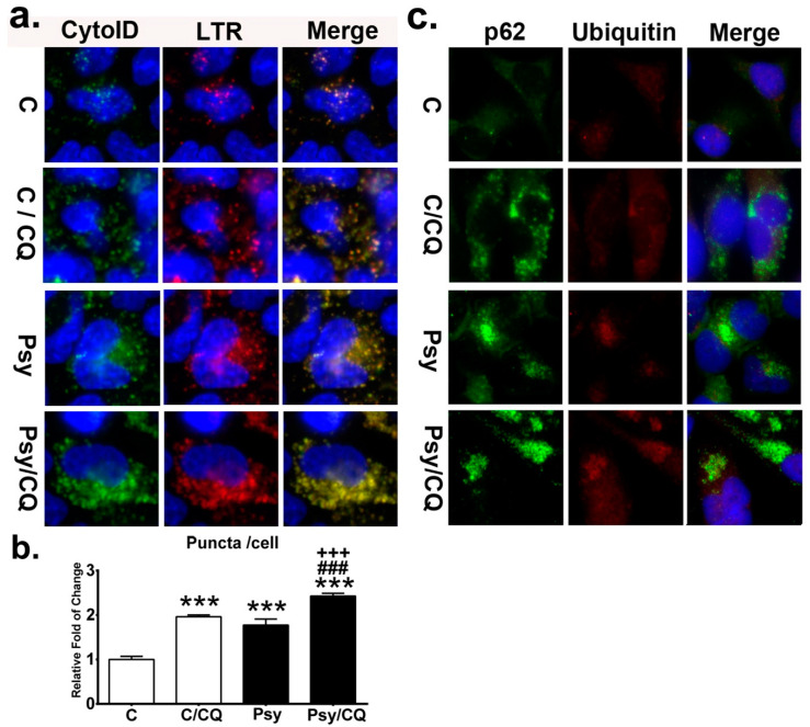
Figure 9.
Accumulation of autolysosomes and p62 aggregates by impaired autophagic flux. (a) Autolysosome was determined by the colocalization of the Cyto-ID fluorescence and LysoTracker Red (LTR) in MO3.13 cells treated with/without psychosine (Psy) concomitant with/without chloroquine (CQ) and counter-stained with Hoechst. (b) Quantification of autolysosome puncta in cells. (c) Cytoplasmic deposition of p62 and ubiquitin by chloroquine. Cells were double labeled with anti-p62 and anti-ubiquitin and counterstained with DAPI. Values are expressed as mean ± S.D. (n = 3). Statistical significance *** p< 0.001 in comparison to control (C). C/CQ vs. Psy, or Psy/CQ, ###p < 0.001. Psy vs. Psy/CQ, +++p < 0.001.

