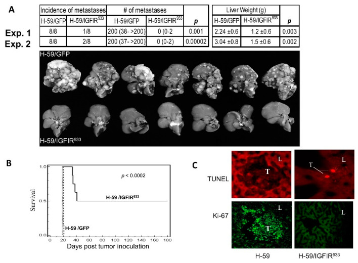
Figure 1.
Loss of metastatic potential in lung carcinoma cells expressing a soluble IGF-IR decoy (IGFIR933). Lewis lung carcinoma subline H-59 cells were transduced with retroparticles expressing the truncated 933 aa IGF-IR decoy (H-59/IGFIR933) or GFP only (H-59/GFP) and 105 tumor cells injected into syngeneic C57Bl/6 female mice via the intrasplenic/portal route to generate experimental liver metastases. Mice were sacrificed and visible metastases enumerated 14 days later. Shown in (A) (top) are the median numbers of metastases (and range) per liver based on eight animals per group in two separate experiments. Liver weights (means ± SD) are shown on the right, and representative livers from experiment (Exp.) 2 are shown on the bottom. Shown in (B) are survival data for mice inoculated in a similar manner (p < 0.0002) and in (C) terminal deoxynucleotidyl transferase (Tdt)-mediated nick end labeling (TUNEL) assay (top) and Ki-67 staining (bottom) performed on liver (L) cryostat sections prepared 5 days post tumor (T) injection (Mag. X135). Reproduced from [88].

