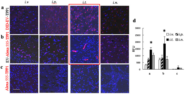
Figure 3.
Brain distribution of fluorescently labeled (a,b) EV-TPP1 formulations and (c) TPP1 alone in BD mice. CLN2 KO mice (1 month old, N = 4) were administered with: (a) DID-EVs (red) loaded with non-labeled TPP1, or (b) non-labeled EVs loaded with fluorescently labeled Alexa 555-TPP1 (red), or (c) Alexa 555-TPP1 alone without EVs (red) through i.v. (2 × 10¹⁰ particles/200 µL), or i.p. (2 × 10¹⁰ particles/200 µL), or i.t. (5 × 10⁹ particles/50 µL), or i.n. (2 × 10⁹ particles/20 µL) routs. 72 h later, mice were sacrificed, perfused, brain slides were processed, and examined by confocal microscopy. Nuclei were stained with DAPI (blue). Obtained confocal images (a–c), and quantification of fluorescence signals (d) indicate the significant accumulation of EVs nanocarriers (a,d), as well as TPP1 in EVs (b,d) in the mouse brain for all administration routes, especially after i.t. injection. Little, if any, TPP1 fluorescence was found in the brain when TPP1 was administered alone without EVs nanocarriers (c, d). The bar: 50 µm. Values are the means ± SEM, * p < 0.05.

