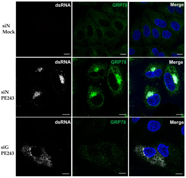
Figure 5.
Viral dsRNA localisation is altered following GRP78 depletion. A549 cells were treated with siN or siG and mock infected or infected with ZIKV PE243 at MOI 0.1 for 24 h. Cells were stained with an anti-dsRNA antibody (white) and an anti-GRP78 antibody (green), and the nuclei were stained with DAPI (blue). Images were taken on an LSM 710 confocal microscope. Scale bars represent 10 µm, and images are representative of triplicate experiments.

