Figure 3.
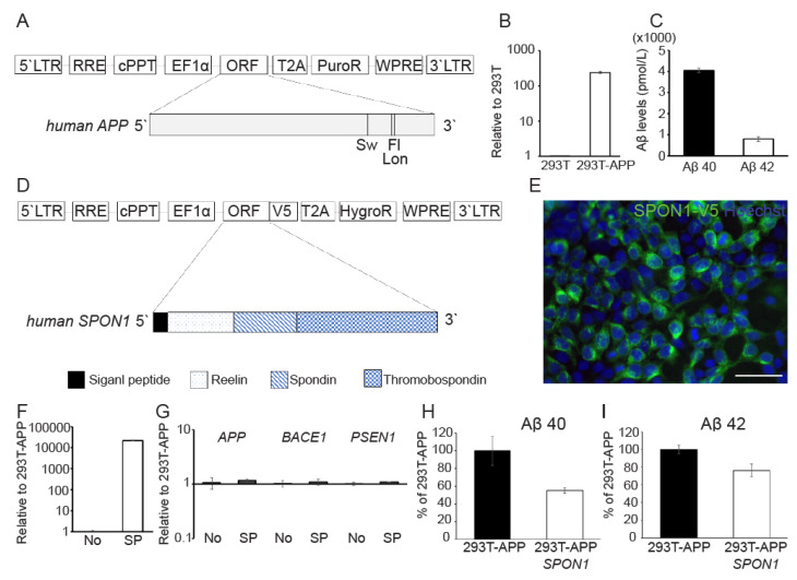
The reduction of Aβ by direct human SPON1 gene delivery. (A) Schematic drawing of lentiviral vector encoding the human APP transgene with Swedish (K670N/M671L), Florida (I716V), and London (V717I) mutations. (B) Quantification of exogenous APP expression in 293T-APP using qRT-PCR. Data are presented as mean ± SEM (n = 3). (C) The quantification of secreted Aβ 40 and Aβ 42 in 293T-APP cultured medium by ELISA. Data are presented as mean ± SEM (n = 3). (D) Schematic drawing of lentiviral vector encoding full length of human SPON1 coding sequences. V5 tag was linked to open reading frame (ORF). (E) Immunofluorescence image of ectopic expression of human SPON1 in 293T-APP-SPON1. Anti-V5 antibody was used to detect V5-tagged SPON1. Scale bar: 50 μm (F) Quantification of exogenous SPON1 expression in 293T-APP (No) and 293T-APP-SPON1 (SP) using qRT-PCR. Data are presented as mean ± SEM (n = 3). (G) The expression of genes related to the amyloidogenic pathway in 293T-APP (No) and 293T-APP-SPON1 (SP) by qRT-PCR. Data are presented as mean ± SEM (n = 3). (H,I) The quantification of secreted Aβ 40 (H) and Aβ 42 (I) in 293T-APP and 293T-APP-SPON1 cultured medium by ELISA. The amount of secreted Aβ 40 or Aβ 42 was normalized to 100% in 293T-APP. Data are presented as mean ± SEM (n = 3).

