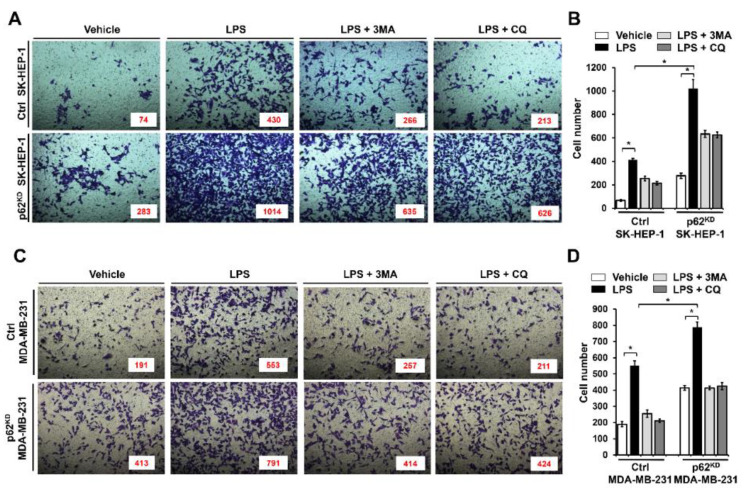
Figure 5.
p62KD SK-HEP-1 and p62KD MDA-MB-231 cells exhibit increased invasiveness in response to TLR4 stimulation. (A,B) Ctrl and p62KD SK-HEP-1 cells were suspended in DMEM culture medium including vehicle, LPS (10 μg/mL), 3-MA (5 mM) plus LPS (10 μg/mL), and CQ (10 μM) plus LPS (10 μg/mL). Cells were placed into the top chambers of 24-transwell plates and incubated for overnight. Fixed cells were stained by using crystal violet (A). Numbers of migrated cells were counted, and results are represented as mean ± SEM (B). * p < 0.05. (C,D) Ctrl and p62KD MDA-MB-231 cells were suspended in culture medium of RPMI including vehicle, LPS (10 μg/mL), 3-MA (5 mM) plus LPS (10 μg/mL), and CQ (10 μM) plus LPS (10 μg/mL). Cells were placed into top chambers of 24-transwell plates and further incubated for overnight. Fixed cells were stained with crystal violet (C). Numbers of migrated cells were counted, and results are represented as mean ± SEM (D). * p < 0.05.

