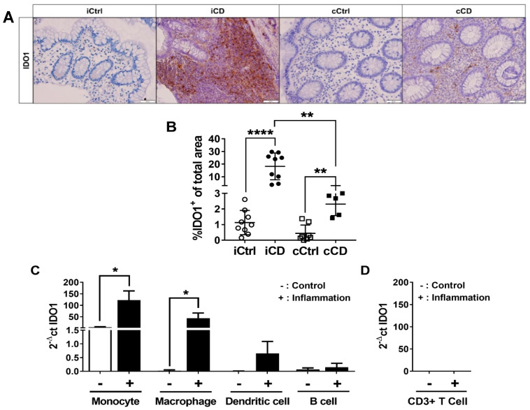
Figure 2.
IDO1 expression is increased in the intestinal tissue of patients with active CD. (A) Analysis of IDO1 protein expression in paraffin-embedded tissue samples of patients with CD using IHC (representative images, magnification 20×). Inflamed tissue of patients with CD (ileum and colon) were compared to noninflamed control samples. Active inflammation was determined using the Riley score. (B) Differential quantitative and localization-dependent evaluation of total IDO1+ area in patients with ileal and colonic CD, compared to control samples. (C,D) Distribution and regulation of IDO1 mRNA expression in (C) Antigen-presenting cells and (D) CD3+ T cells. Antigen-presenting cells and CD3+ T cells were stimulated for 24 h with IFNγ (100 U/mL), TNFα (100 ng/mL) and IL-1β (10 ng/mL). mRNA expression levels were determined by TaqMan real-time PCR as described in Section 2. (B–D) Data are shown as mean ± SD of n = 3–9; * for p ≤ 0.05, ** for p < 0.01, and **** for p < 0.0001 using the (B) Mann–Whitney test or (C,D) paired Student’s t-test. cCD = colonic CD, cCtrl = colon non-IBD control, iCD = ileal CD, iCtrl = ileum non-IBD control.

