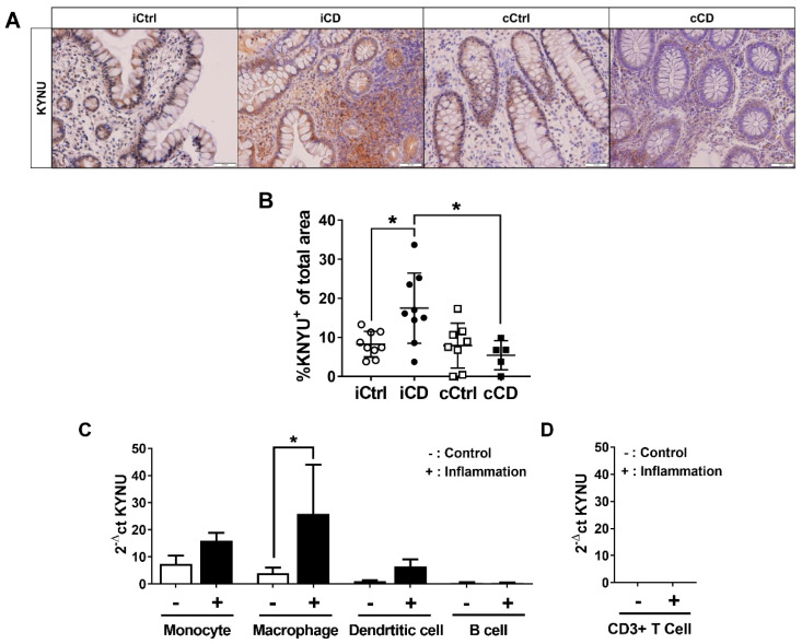
Figure 4.
Differential kynureninase (KYNU) expression in tissue samples of patients with ileal and colonic CD. (A) Analysis of KYNU protein expression in paraffin-embedded tissue of CD patients via IHC (representative images, magnification 20×). Inflamed tissue of patients with ileal and colonic CD were compared to noninflamed control samples from the respective bowel segments. Active inflammation was determined by the Riley score. (B) Differential quantitative and localization-dependent evaluation of total KYNU+ area in CD, compared to non-IBD control (C,D) Distribution and regulation of KYNU mRNA expression in (C) antigen-presenting cells and (D) CD3+ T cells (no signal detected). Antigen-presenting cells and CD3+ T cells were stimulated for 24 h with IFNγ (100 U/mL), TNFα (100 ng/mL) and IL-1β (10 ng/mL). mRNA expression levels were determined by TaqMan real-time PCR as described in Section 2. (B–D) Data are shown as mean ± SD of n = 3–9; * for p ≤ 0.05 using the (B) Mann–Whitney test or (C,D) paired Student’s t-test. cCD = colonic CD, cCtrl = colon non-IBD control, iCD = ileal CD, iCtrl = ileum non-IBD control.

