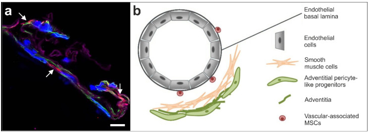
Figure 1.
(a) Immunofluorescent detection of NG2 (red; arrows), smooth muscle antigen (SMA) (green) and laminin (purple) in the wall of a small arteriole; cell nuclei are in blue (modified from [3]). The scale bar represents 10 µm. (b) Schematic illustration of the hypothesized location of vascular associated MSCs (taken from [3] and modified from [35]).

