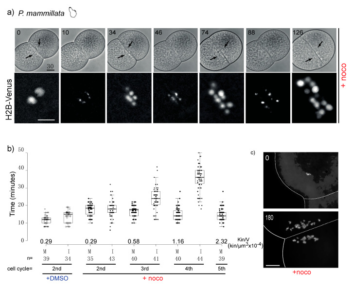
Figure 3.
P. mammillata 2-cell embryos do not arrest in mitosis in the absence of spindle microtubules. (a) Selected frames from a time-lapse movie of a P. mammillata embryo expressing the DNA reporter, H2B-Venus, treated with 10 µM nocodazole after first cleavage. Numbers indicate minutes after treatment. Arrows indicate nuclei visible in bright field optics. See movie 1. (b) Duration of mitosis (M, NEB to NER) and interphase (I, NER to NEB) in P. mammillata embryos treated with DMSO or nocodazole. Kin/V indicates kinetochore to cell volume ratio at each cell cycle starting from fertilization (1st is 1-cell mitosis; 2nd is 2-cell mitosis). Box plot parameters as in Figure 2b. In nocodazole-treated embryos the amount of DNA increases due to subsequent rounds of DNA replication without intervening cytokinesis. Duration of mitosis in control DMSO-treated embryos is constant in all four analyzed cell cycles. (c) DAPI (4′,6-diamidine-2′-phenylindole dihydrochloride)-stained chromosome spreads from DMSO and nocodazole-treated (180 min) P. mammillata embryos. After 180 min nocodazole treatment, embryos have undergone 4 more cell cycles and have 4 times more DNA than at time 0. In the absence of microtubules, the nuclei which form around chromosome clusters (karyomeres) do not fuse and are dispersed over time by cytoplasmic flow (in (a,c)). Scale bar = 30 µm.

