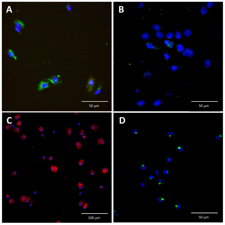
Figure 2.
Immunofluorescence staining for interleukin-6 in liver sinusoidal endothelial cells and Kupffer cells. (A): Isolated liver sinusoidal endothelial cells (LSEC) were stained for stabilin-2 (green), and their nuclei were stained with Hoechst (blue). (B): In the absence of stimulation, IL-6 was not detected in LSEC (not shown). In contrast, after incubation of LSEC with APR, IL-6 (green) was detected in vesicles, with a homogeneous distribution in the cytoplasm. (C): Isolated Kupffer cells (KC) were stained for IBA-1 (red) and their nuclei were stained with Hoechst (blue). (D): KC were stimulated with lipopolysaccharide (0.5 mg/mL) and exhibited a different cytoplasmic distribution of IL-6 than LSEC. In LSEC, IL-6 was more homogeneously distributed in the cytoplasm (B) around the nuclei, whereas in KC, IL-6 appeared in clustered vesicles (D).

