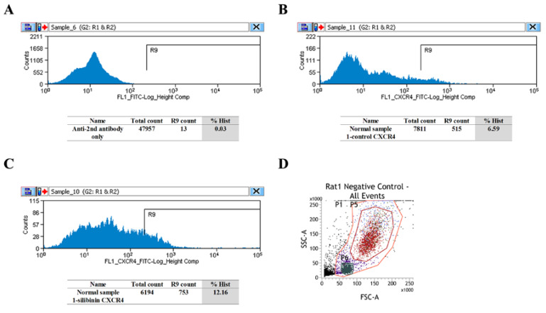
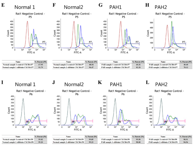
Figure 3.
Flow cytometry (FCM) evaluation of CXCR4 in normal and PAH rats. (A–C) In the BM of normal rats, silibinin upregulated the percentage of CXCR4-positive cells. Secondary antibody only (A), normal 1-control (B), and normal 1-silibinin (C). (D) Negative control of granulocytes (P5 area, red) and negative control of monocyte-macrophages (P6 area, dark green). (E–H) In the BM of normal and PAH rats, silibinin increased the percentage of CXCR4-positive cells in the granulocyte fraction. Negative control (red), normal 1-control (light green), and normal 1-silibinin (blue) (E). Negative control (red), normal 2-control (light green), and normal 2-silibinin (blue) (F). Negative control (red), PAH 1-control (light green), and PAH 1-silibinin (blue) (G). Negative control (red), PAH 2-control (light green), and PAH 2-silibinin (blue) (H). (I–L) In the BM of normal and PAH rats, the percentage of CXCR4-positive cells was not altered in the monocyte-macrophage fraction. Negative control (dark green), normal 1-control (light green), and normal 1-silibinin (blue) (I). Negative control (dark green), normal 2-control (light green), and normal 2-silibinin (blue) (J). Negative control (dark green), PAH 1-control (light green), and PAH 1-silibinin (blue) (K). Negative control (dark green), PAH 2-control (light green), and PAH 2-silibinin (blue) (L). Treatment groups consisted of two samples in each culture.


