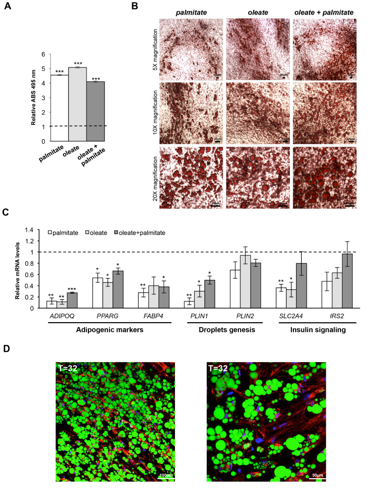
Figure 3.
Hypertrophic-like cells generated from hMSCs-derived adipocytes. (A) Optical determination of lipid accumulation (Oil Red O staining) in hypertrophic-like adipose cells (HAs) generated from mature adipocytes (MAs)—differentiated in vitro by hMSCs—by supplementation of three different fatty acids mixes. Data are shown as mean ±SEM compared to mature adipocytes from three independent experiments. ***p val ≤ 0.001. (B) Representative bright-field images of HAs—generated by three different fatty acids mixes—after lipid droplets staining by Oil Red O (scale bar, 50 µm). (C) Relative mRNA quantification (qPCR) of PPARG and key target genes in HAs generated by three different treatments. PPIA was used as reference gene. Data are reported as mean ±SEM vs. mature adipocytes (dotted line) from three independent experiments. * p val ≤ 0.05, ** p val ≤ 0.01 and *** p val ≤ 0.001. (D) Representative confocal microscopy images of HAs (T = 32d) generated by palmitate-containing mix. Nuclei were stained by DAPI (blue), lipid droplets, and cell membranes by Bodipy 495/503 (green) and WGA 632/647 (red), respectively (scale bar, 100 µm left panel; scale bar, 50 µm right panel).

