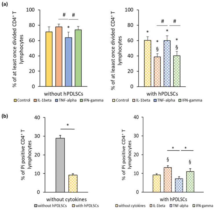
Figure 1.
hPDLSC mediated effect of different inflammatory stimuli (IL-1β, TNF-α, and IFN-γ) on CD4+ T lymphocyte proliferation and apoptosis. Primary hPDLSCs were stimulated with either 5 ng/ml IL-1β or 10 ng/ml TNF-α or 100 ng/ml IFN-γ. After 48 h, hPDLSCs were applied to indirect co-culture and stimulated as described above. Allogenic CD4+ T lymphocytes were added to indirect co-culture using Transwell (TC) inserts. Additionally, CD4+ T lymphocytes were cultured in the absence of hPDLSCs and stimulated with either 5 ng/ml IL-1β or 10 ng/ml TNF-α or 100 ng/ml IFN-γ. CD4+ T lymphocyte proliferation was induced by 10 µg/ml phytohemagglutinin-L (PHA-L). After five days incubation, CD4+ T lymphocyte proliferation (a) was determined by flow cytometry by analyzing carboxyfluorescein succinimidyl ester (CFSE) labeled CD4+ T lymphocytes. The Y-axis shows the percentage of at least once divided CD4+ T lymphocytes. Additionally, after five days incubation, the percentage of apoptotic CD4+ T lymphocytes (b) was determined by flow cytometry by labeling CD4+ T lymphocytes with Pi. The Y-axis shows the percentage of Pi positive CD4+ T lymphocytes. Data are presented as mean value ± S.E.M. received from five independent experiments with hPDLSCs from five different individuals. * p-value < 0.05 compared to the control (without hPDLSCs); § p-value < 0.05 compared to CD4+ T lymphocytes in the presence of untriggered hPDLSCs; # p-value < 0.05 compared between groups as indicated.

