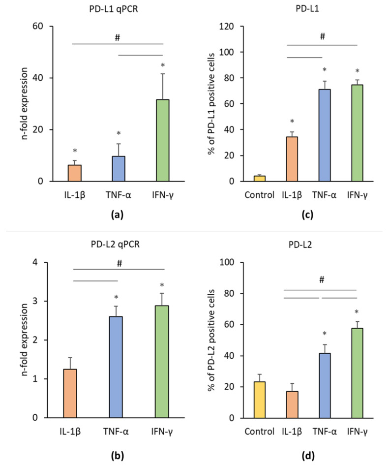
Figure 3.
Effect of pro-inflammatory stimuli (IL-1β, TNF-α, and IFN-γ) on programmed cell death 1 ligand 1 (PD-L1) and programmed cell death 1 ligand 2 (PD-L2) production in hPDLSCs. Primary hPDLSCs were treated with either 5 ng/ml IL-1β or 10 ng/ml TNF-α or 100 ng/ml IFN-γ for 48 h. Unstimulated hPDLSCs served as control. PD-L1 (a) and PD-L2 (b) gene expression levels were determined by qPCR, demonstrating the n-fold PD-L1 / PD-L2 expression compared to the control (n = 1). GAPDH served as internal reference gene. PD-L1/PD-L2 protein levels were investigated by intracellular immunostaining followed by flow cytometry analysis, determining the percentage of PD-L1 (c) and PD-L2 (d) positive cells. All data are presented as mean value ± S.E.M. received from six independent experiments with cells isolated from six different individuals. * p-value < 0.05 compared to the unstimulated control; # p-value < 0.05 compared between groups as indicated.

