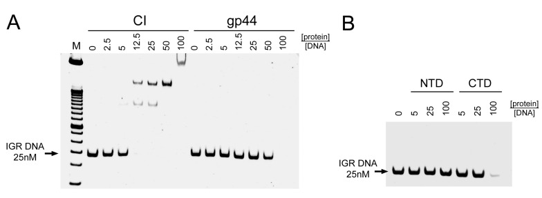
Figure 4.
DNA binding of gp44 and CI. (A) Separation on a 6% polyacrylamide DNA retardation gel of 25 nM intergenic region (IGR) DNA after incubation with varying concentrations of CI (left) or gp44 (right). (B) IGR DNA incubated with gp44-NTD or gp44-CTD. The protein concentrations are given above the gel as the -fold molar excess of protein to DNA. The position of the IGR DNA band is indicated.

