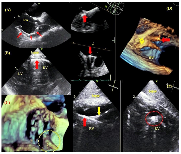
Figure 3.
Binding sites between leads and cardiovascular structures visualized by TEE. TEE—bicaval view—a thickened lead (red arrows) adhered to the wall of the right atrium and the superior vena cava orifice (A). TEE—transgastric view—a ventricular lead (arrow) adhered to the right ventricular wall (B). 3D TEE imaging—the tricuspid valve with a lead adhered to the leaflet margin (C). 3D imaging (Multi-D)—a lead (red arrow) implanted at the base of the papillary muscle (yellow arrow) (D). TEE (Multi-D)—a ventricular lead (red arrow) adhered to the tendinous thread (yellow arrow) (E).

