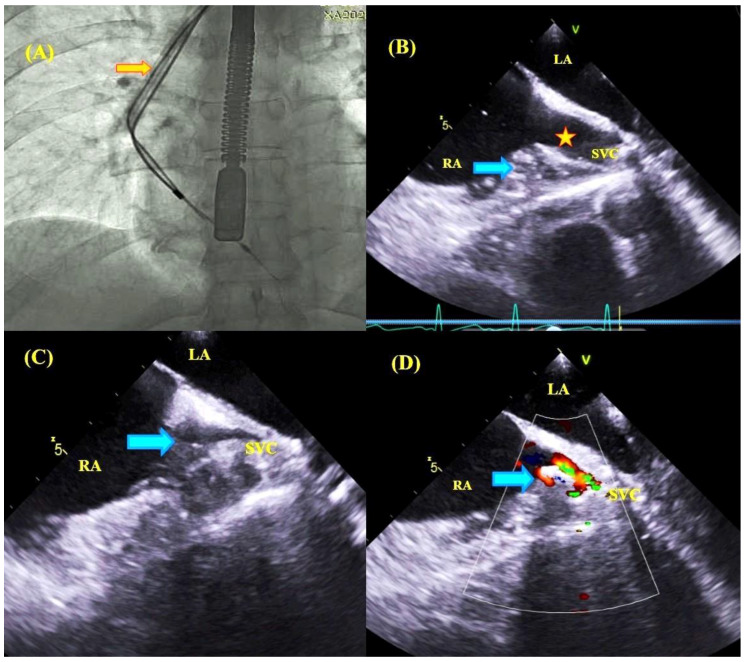
Figure 5.
Comparison of fluoroscopic and TEE images during removal of lead-to-lead adhesions. Fluoroscopic image—the moment of ventricular lead extraction with simultaneous pulling on the atrial lead; the Byrd catheter slipped over the ventricular lead (orange arrow) (A). TEE images—mid-esophageal view, consecutive phases of pulling on the atrial wall and superior vena cava (blue arrows) until marked obliteration of the vessel during ventricular lead extraction and pulling on atrial lead adhesion (B–D).

