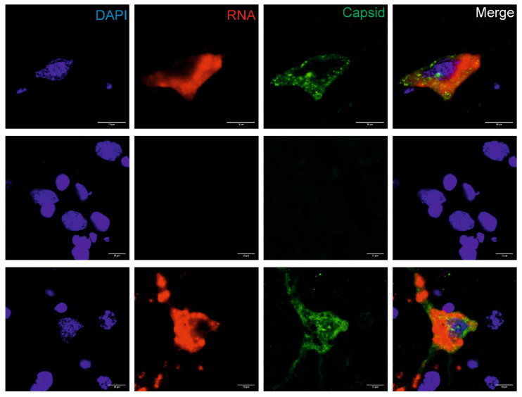
Figure 3.
In situ detection of BoAstV CH13 in the hippocampus. Formalin-fixed, paraffin-embedded (FFPE) brain sections of the hippocampus of the BoAstV PE3373/2019/Italy case (top panel), a negative control animal (middle lane), and a BoAstV CH13 positive case (bottom panel) are presented. Cell nuclei were stained with DAPI (blue), viral RNA by fluorescent in situ hybridization (red), and the viral capsid protein by immunofluorescence (green). In the BoAstV PE3373/2019/Italy case and in the positive control, viral RNA and antigens co-localized the same cells (merged pictures), which suggests active replication of the virus. The negative control shows neither staining for viral RNA, nor for viral antigen. Microphotographs were taken at ×60 magnification.

