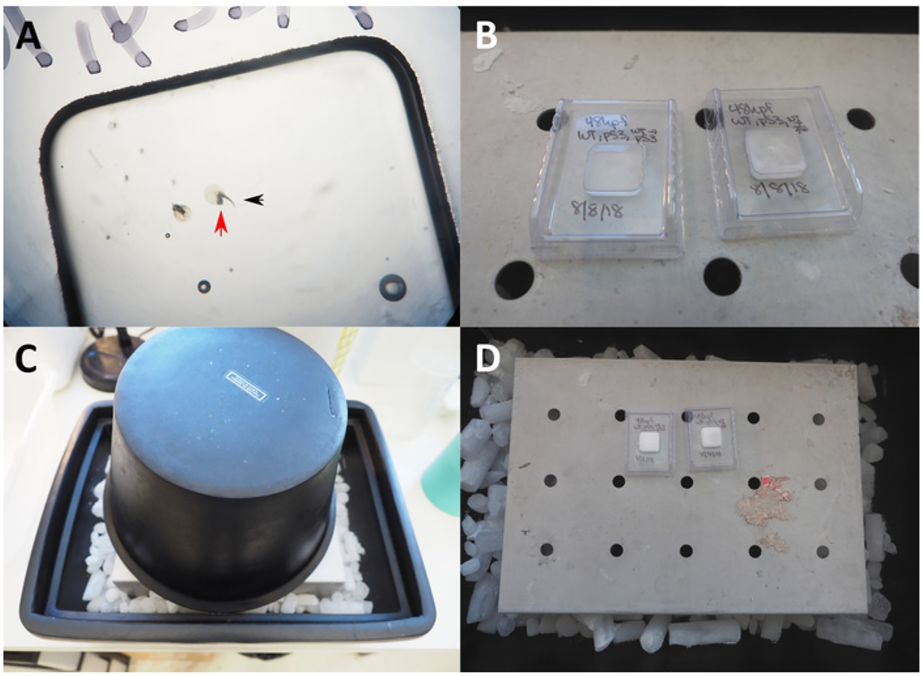
Figure 1: Preparation of frozen OCT blocks.

(A) 50x microscopic view of embryos in OCT in a cryogenic mold prepared for freezing. Red arrow indicates head of zebrafish embryo position against the bottom surface of the mold; black arrow indicates tail pointing up towards the user. (B) Plastic molds with embryos and OCT placed on chilled metal platform. (C) Foam ice bucket placed over plastic molds to create freezing chamber. (D) OCT turns white from transparent once the block is fully frozen.
