Figure 3.
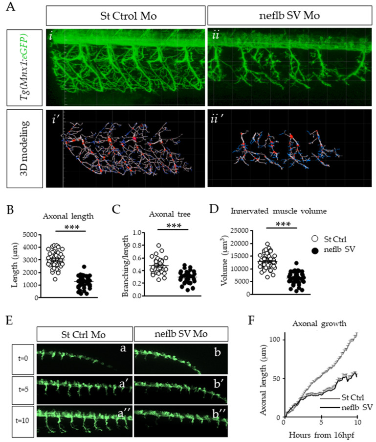
Axonal atrophy in motor neurons of neflb SV embryos. (A), in vivo observation of somitic nerve fascicles in 48 hpf Hb9:eGFP zebrafish embryos injected with a control Mo (i) or with neflb SV Mo (ii). Hb9:eGFP line express the GFP (green) in motor neurons under the specific promoter HB9. After the neflb SV Mo injection, the axonal projections appeared shorter and less regularly distributed and branched than controls. (i’,ii’), 3D modeling of motor neuron morphology using the Imaris software. This reconstruction permitted the quantitative analysis of the nerve fascicles features: (B), the axonal length; (C), the axonal branching; (D), the volume of the innervated muscle. All measurements were reduced in the neflb SV morphants (black circles) with respect to the controls (white circles). (E), in vivo time-lapse imaging of the Tg(Mnx1:eGFP) zebrafish embryos injected with a control morpholino (a–a″) or with the neflb SV morpholino (b–b″) was performed during 10 h, starting from 16 hpf. (F), quantification of motor neurons axonal length during time. hpf, hour post fertilization. *** p < 0.001.

