Figure 2.
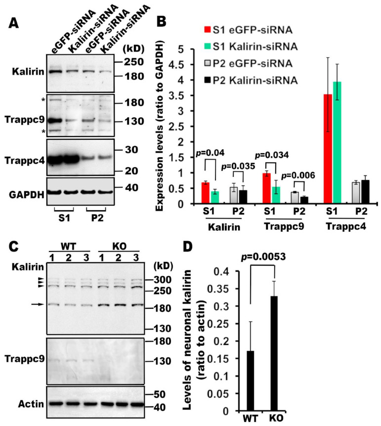
Kalirin and trappc9 mutually affect their expression. (A) and (B) Kalirin knockdown perturbs the expression as well as membrane association of trappc9. (A) Post-nuclear supernatants (S1) and cellular membranes (P2) from NRK cells treated with siRNAs for eGFP or kalirin were analyzed by Western blot with indicated antibodies. Shown are blot analyses from one of three experiments with similar results. Note that the blots were re-probed with anti-trappc9 antibodies after incubation with anti-kalirin antibodies, therefore signals detected by anti-kalirin antibodies still persisted as indicated by stars (*). (B) Films with signals in linear ranges were scanned into digital images for measuring intensities of signals for each protein and corresponding background signals using NIH ImageJ. Data are expressed as ratio of signal intensity of the indicated protein to that of GAPDH (n = 3, Mean ± SD, two-tailed Student’s t-test). (C) and (D) Constitutive loss of trappc9 causes elevated expression of kalirin in brain neurons. (C) Western blot analysis of post-nuclear supernatants from brain tissues of three wild-type (WT) and three trappc9 knockout (KO) mice. The genotype of all mice was determined by PCR. The age of all mice used for harvesting brain tissues was 3 months. The arrow points to the neuronal isoform of kalirin (190 kD), whereas arrowheads indicate other kalirin isoforms. (D) Intensities of signals for the neuronal kalirin isoform as well as actin and background in the same lane were measured with NIH ImageJ as above and used for calculating Mean ± SD ratio of neuronal kalirin to actin signals. Comparison was performed with two-tailed Student’s t-test.

