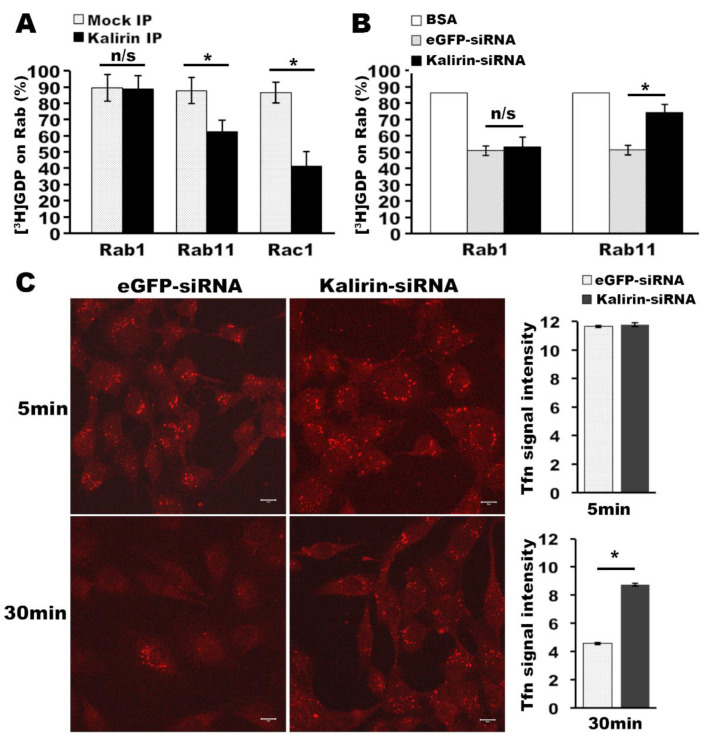
Figure 3.
Kalirin regulates the activation of Rab11 and endocytic recycling of transferrin. (A) Kalirin immunoprecipitates containing GEF activities on Rab11. Triton solubilized cellular membranes from cells transfected with pEAK-His-myc-kalirin7 or empty vector were used for immunoprecipitation with anti-myc antibodies. Precipitated proteins were used for the [3H]GDP release assay as described in Methods. Mean ± SD percentages of [3H]GDP remaining on GST-Rab1-His, GST-Rab11-His, and GST-Rac1 were graphed (n = 3, two-tailed Student’s t-test: n/s, not significant, * p < 0.01). (B) Knockdown of kalirin reduced Rab11GEF activities in cellular membranes. After knocking down kalirin in NRK cells as in (2A), cellular membranes were prepared and extracted in GEF assay buffer containing triton X-100. Equal amounts of solubilized membranes from cells treated with eGFP-siRNA and kalirin-siRNA were used for [3H]GDP release from GST-Rab1-His and GST-Rab11-His as above. BSA is a no-GEF control (n = 3, Mean ± SD, two-tailed Student’s t-test: n/s, not significant, * p < 0.01). (C) Synchronized uptake and recycling of Alexa568-transferrin was performed with siRNA-treated NRK cells for indicated times. After uptake, NRK cells on glass coverslips were processed for fluorescent microscopy. Shown are confocal images. Scale bar: 10 μm. Signal intensities of Alexa568-transferrin and the corresponding background of the images for each condition were measured using NIH ImageJ. After subtracting the background, the mean values of Alexa568-transferrin signals were calculated and plotted (n = 33 cells for kalirin-siRNA and 34 cells for eGFP-siRNA for 5 min incubation, n = 35 cells for kalirin-siRNA and 31 cells for eGFP-siRNA for 30min incubation; Mean±SD, two-tailed Student’s t-test: * p < 0.01).

